Why we should use Köhler illumination
Köhler illumination is an essential technique for setting up any widefield microscope and adjusting it properly.
The downloadable PDF files on the right give instructions on how to set up, and properly align, any light microscope according to Köhler’s principles of illumination.
Nowadays we expect the specimen to be illuminated uniformly over the entire field of view. However, prior to the widespread use of electric light, this could be accomplished only by using a cloudy sky as the light source. This simple type of illumination is called ‘source-focused’ or ‘critical’ illumination. Electric lamps were introduced into microscopy towards the end of the 19th century, and provided a clean, reliable, intense and immediate source of illumination. They were cleaner and safer to use that oil or gas illumination, and more reliable than depending upon suitable solar illumination. However, the electric lamp filament possesses a definite structure which, if imaged onto the specimen by source-focused illumination, will interfere with interpretation of the image. In 1893 August Köhler devised the method of illumination bearing his name to circumvent this problem.
A comprehensive explanation of the principles of Köhler illumination has been written by Dr. Peter Evennett, made available by permission of the Royal Microscopical Society. Download it from the sidebar on the right. There is also a related article by Dr. Evennett explaining Depth of field and Depth of Focus, and the difference between these two terms. A further information sheet that I have written on how to adjust the eyepieces correctly, after the microscope has been adjusted for Köhler illumination, is also available for download. The topic of Köhler illumination, light sources and illumination in the microscope is covered in Chapter 9 of Understanding Light Microscopy.
Advantages of Köhler illumination
- Provides a large and uniform, intense, structureless light source from a small, filament structure
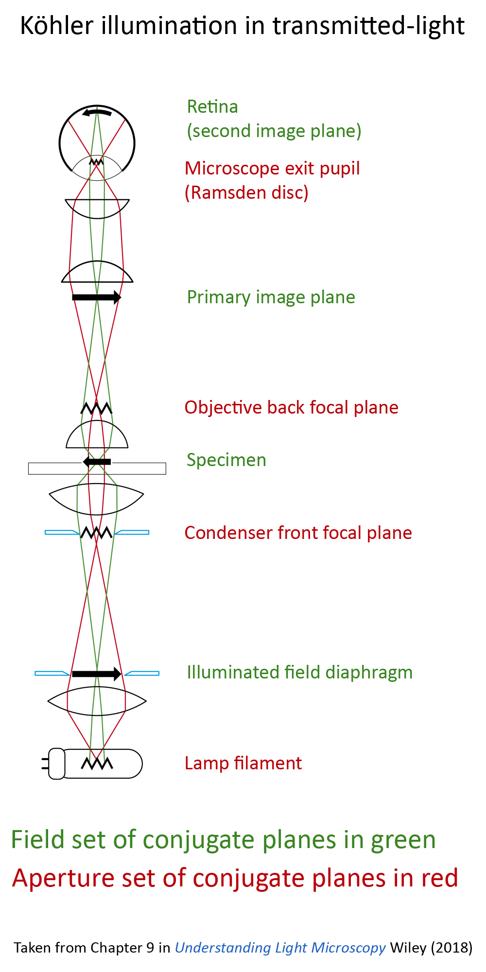
- The illuminating filament in the aperture set of conjugate planes does not disturb the image which lies in the field set of conjugate planes
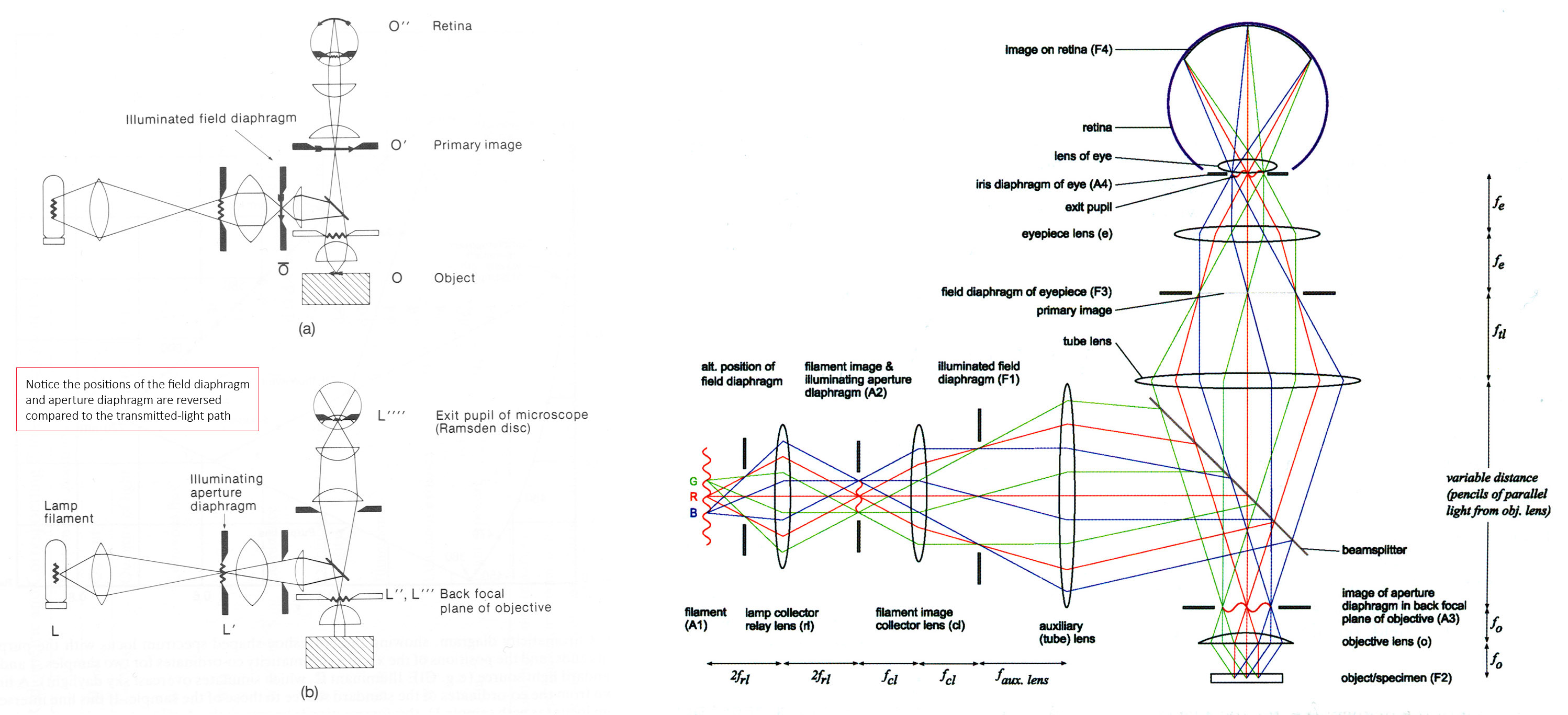
- Kohler illumination allows independent control of the illuminating and imaging ray paths.
- Stray non image-forming light can (largely) be excluded from the microscope, leading to improved contrast.
- Rays arising from each individual point on the filament pass through all parts of the object
- Each individual point on the object receives light from all parts of the filament admitted by the condenser.
The image of the lamp collector lens, bounded by the illuminated field diaphragm, provides a flawless field of uniform illumination. The illuminated field diaphragm acts as the control for area of illumination. The illuminating aperture diaphragm (condenser diaphragm) controls the included angle of illumination focused by the condenser onto the object.
References
-
- Evennett, PJ (1983) Köhler illumination: A simple interpretation Proc. RMS 28/4: 189-192
- Evennett, PJ et al (2009) Symmetrical ray diagrams of the optical pathways in light microscopes Quekett Journal of Microscopy 41/2: 109-118 – infinity-corrected microscope. This paper, Evennett et al (2009) is an update of the (2006) paper written by Hammond & Heath.
- Hammond, C (1998) Symmetrical representation of the geometrical optics of the light microscope Jour. Microscopy 192/1: 63-68 – finite tube-length microscope
- Evennett, PJ (1996) Depth of Field and Depth of Focus Explained Proc. RMS 31/1: 64-66
- Köhler, A (1893) “Ein neues Beleuchtungsverfahren für mikrophotographische Zweckeâ€. Zeitschrift für wissenschaftliche Mikrosk. Mikrosk. Tech., 10(4): 433-440
- Köhler, A (1894) New Method of Illumination for Photomicrographical Purposes Journal of the Royal Microscopical Society. 14: 261-262 – a summary report
A4 sheet of Köhler protocol
Stereo-microscope adjustment instructions (for keeping by the stereo-microscope)
Quick instructions (compound & stereo-microscopes) A4 sheet
Explanation of Köhler illumination (Dr Peter Evennett)
Depth of Field and Depth of Focus Explanation
(Dr Peter Evennett)
Adjustment of the eyepiece ‘dioptre controls’ for parfocality
(Jeremy Sanderson)